Condition Guide

Adult Scoliosis

Spinal curvature in adults progresses with time and can cause pain, disability and neurological problems. Expert surgical correction restores alignment and alleviates symptoms. Here is what you need to know.

Posterior Fusion ALIF/OLIF Minimally Invasive Complex Deformity Navigation-Guided
25+
Years Experience
2
Board Certifications
100+
Deformity Corrections
AOSpine
North America Faculty
Understanding the Condition

What Is Adult Scoliosis?

Adult scoliosis is a sideways curvature of the spine that develops or worsens after skeletal maturity. Unlike adolescent idiopathic scoliosis (which appears in teenagers), adult scoliosis typically results from degenerative changes in the spine — loss of disc height, facet joint arthritis, and vertebral bone loss with age.

The spine has three regions: the cervical (neck), thoracic (mid-back) and lumbar (lower back) spine. Adult scoliosis most commonly affects the lumbar spine, where it can cause significant pain, imbalance, and neurological symptoms.

Adult scoliosis is progressive. Without treatment, curves typically worsen 1-2 degrees per year. As the curve increases, pain intensifies, walking becomes more difficult, and nerve compression can develop. Surgery is the definitive treatment when conservative measures have failed.

Condition at a Glance
Common Affects 10-30% of adults over age 50. More common in women. Prevalence
Progressive Degenerative scoliosis worsens over time without intervention. Average 1-2 degrees per year. Natural History
Correctable Modern fusion techniques achieve 50-70% curve correction with minimal invasiveness. Surgical Outcomes
Patient Education

Understanding Spinal Deformity

Dr. Ganapathy explains scoliosis, adult spinal deformity, when surgery becomes necessary, and the different surgical approaches available to treat these conditions.

Recognise the Signs

Warning Signs of Adult Scoliosis

Adult scoliosis symptoms typically progress gradually. Many patients adapt to changes without realising the spine has become misaligned. If you notice several of these, book an assessment promptly.

🧍
Uneven Shoulder or Hip Height
One shoulder sitting higher than the other, or hips appearing asymmetrical. Body leaning to one side when standing upright.
💔
Lower Back Pain
Chronic pain in the lumbar spine, often worse with standing or walking. May be unilateral (one side) due to facet joint arthritis.
🚶
Worsening Gait and Balance
Walking becomes stiff or asymmetrical. Difficulty standing straight. Frequent falls or need for support when walking.
Leg Pain or Numbness
Radiating pain down the leg (radiculopathy). Numbness or tingling in the buttocks, thigh or foot. Usually one-sided.
🗜️
Shortness of Breath
Fatigue with exertion. Reduced lung capacity due to severe thoracic scoliosis compressing the rib cage.
🛑
Difficulty Walking Long Distances
Claudication symptoms. Able to walk only short distances before needing to sit. Symptoms improve when seated or bending forward.
Getting an Accurate Diagnosis

How We Diagnose Adult Scoliosis

Diagnosis begins with a detailed history and physical examination. Dr. Ganapathy assesses spinal alignment, range of motion, strength, sensation and reflexes. He looks for signs of neurological compromise from nerve compression.

Standing X-rays (anteroposterior and lateral views) are essential to measure the degree of curvature, assess sagittal balance, and evaluate degenerative changes. Full-length spinal X-rays show the relationship between the curve and the body's centre of gravity.

MRI provides detailed information about the spinal cord, nerve roots, discs and ligaments. It reveals stenosis, neural compression and helps determine the levels requiring fusion. CT scans show bony anatomy in detail when surgical planning requires precise bone measurement.

The Cobb angle measures the degree of scoliosis. Sagittal balance — how the spine aligns with gravity — is equally important:

Severe Cobb angle > 50°. Surgery strongly recommended. Cobb > 50°
Moderate Cobb 30-50°. Surgery often recommended depending on symptoms. Cobb 30-50°
Mild Cobb < 30°. Monitor with imaging every 1-2 years. Cobb < 30°
Surgical Treatment Options

Procedures Dr. Ganapathy Performs

The right procedure depends on the degree of curvature, number of levels involved, sagittal balance, and your overall health. All options are discussed in detail at your consultation.

Standard Approach
Posterior Spinal Fusion (PSF)
Multi-level fusion with pedicle screw fixation and rods. The gold standard for adult scoliosis correction. Typically 4+ levels. Effective for both curve correction and restoration of sagittal balance.
Anterior Approach
ALIF — Anterior Lumbar Interbody Fusion
Direct access to the discs through the front of the abdomen. Used to correct lower lumbar curves, restore disc height and interbody support. Often combined with posterior fusion for maximum stability.
Minimally Invasive
OLIF — Oblique Lateral Interbody Fusion
Minimally invasive lateral access to the discs. Smaller incisions, less muscle damage and faster recovery than traditional open surgery. Excellent curve correction without bone grafting.
Hybrid Approach
Combined Anterior and Posterior Fusion
For severe deformity, combining anterior interbody support with posterior instrumentation provides maximum correction, stability and long-term durability. Two-stage or single-stage options available.
Clinical Outcomes

Before & After: Thoracolumbar Scoliosis & Kyphosis Correction

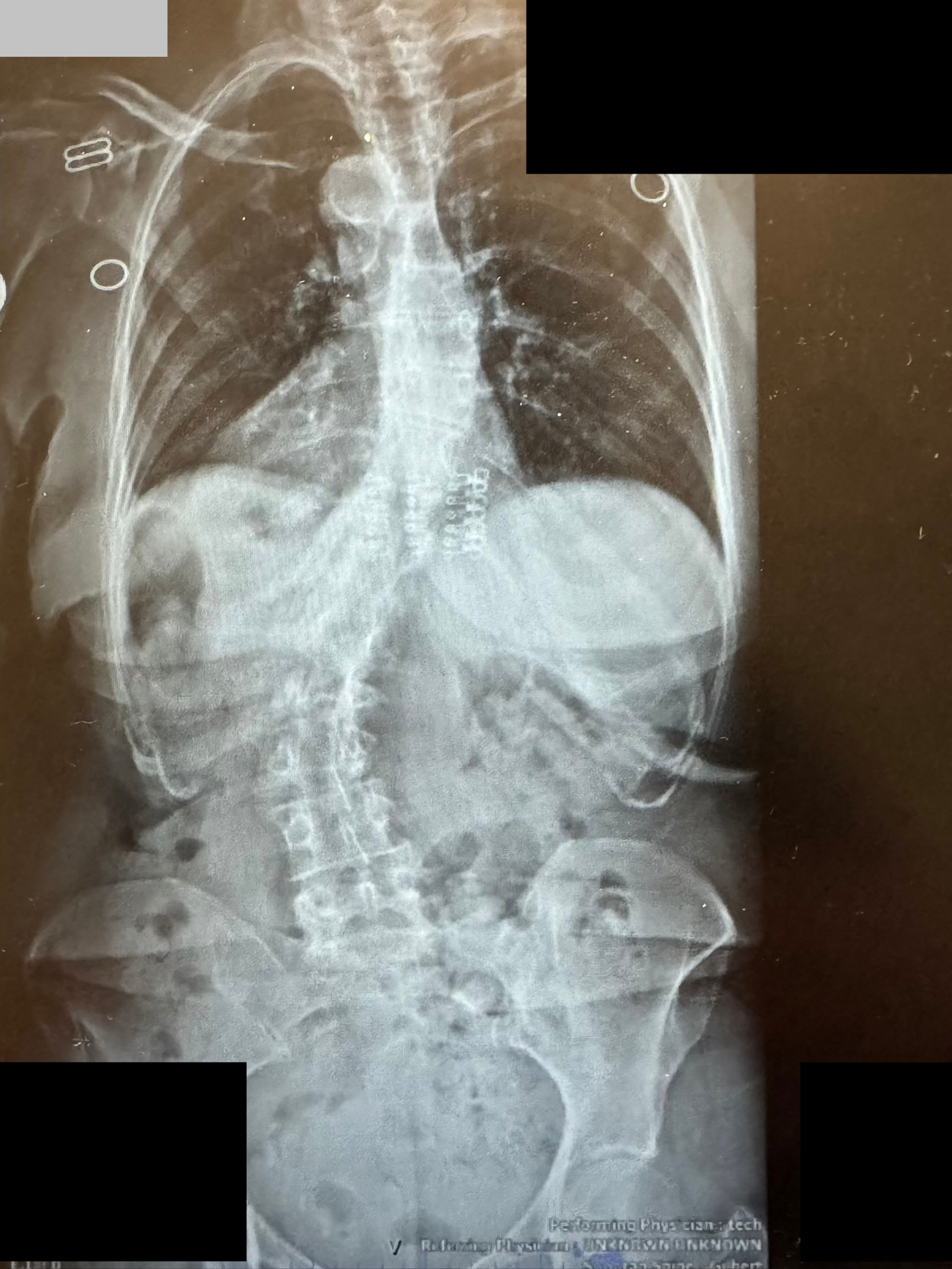
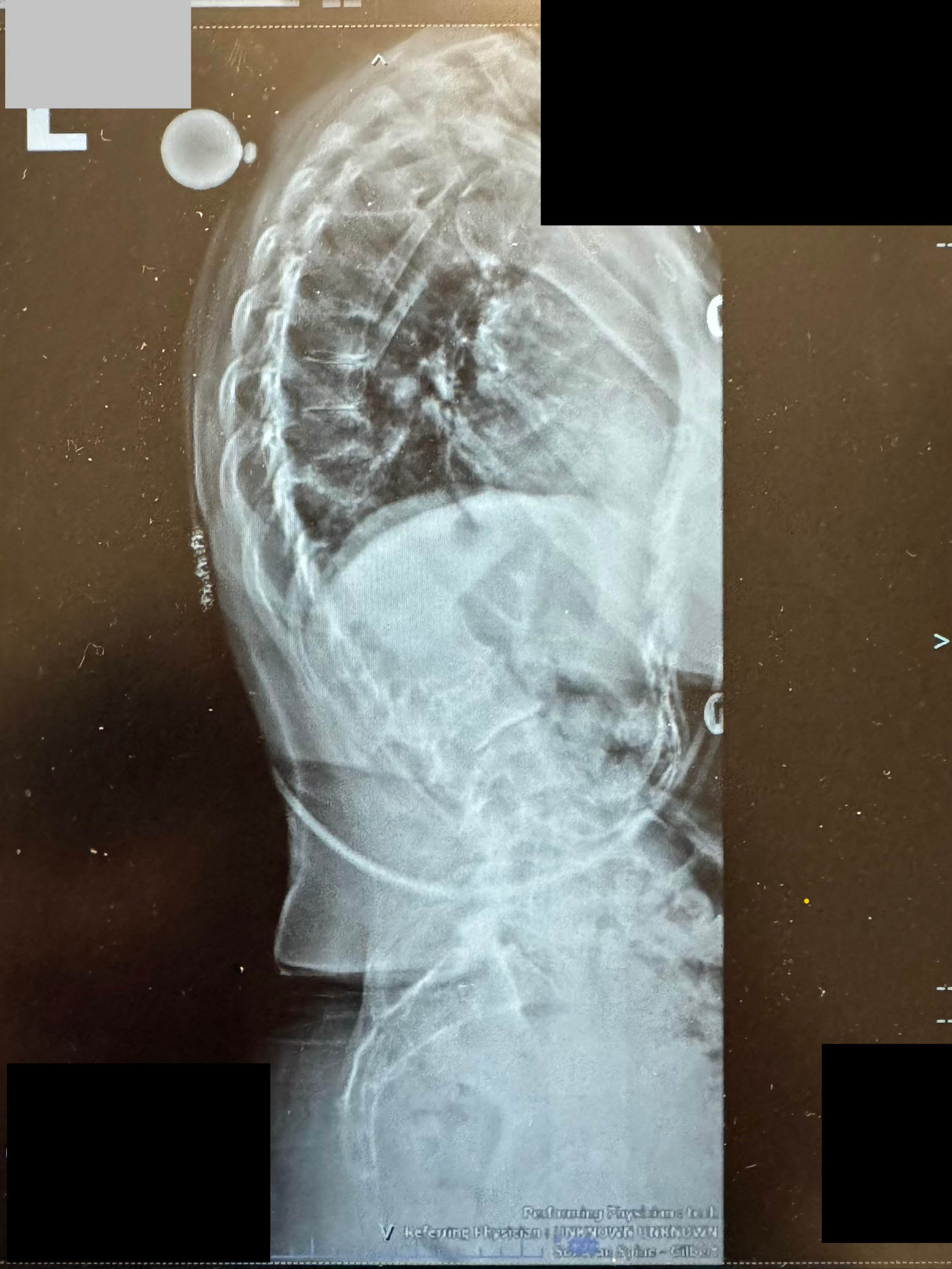
Real patient case showing successful correction of combined thoracolumbar scoliosis and kyphotic deformity using multi-level posterior fusion with instrumentation.

Pre-Operative Imaging

Pre-operative AP X-ray showing thoracolumbar scoliosis deformity
AP View: Significant lateral curve and vertebral rotation visible in thoracolumbar region
Pre-operative lateral X-ray showing excessive kyphosis
Lateral View: Excessive thoracolumbar kyphosis with sagittal imbalance

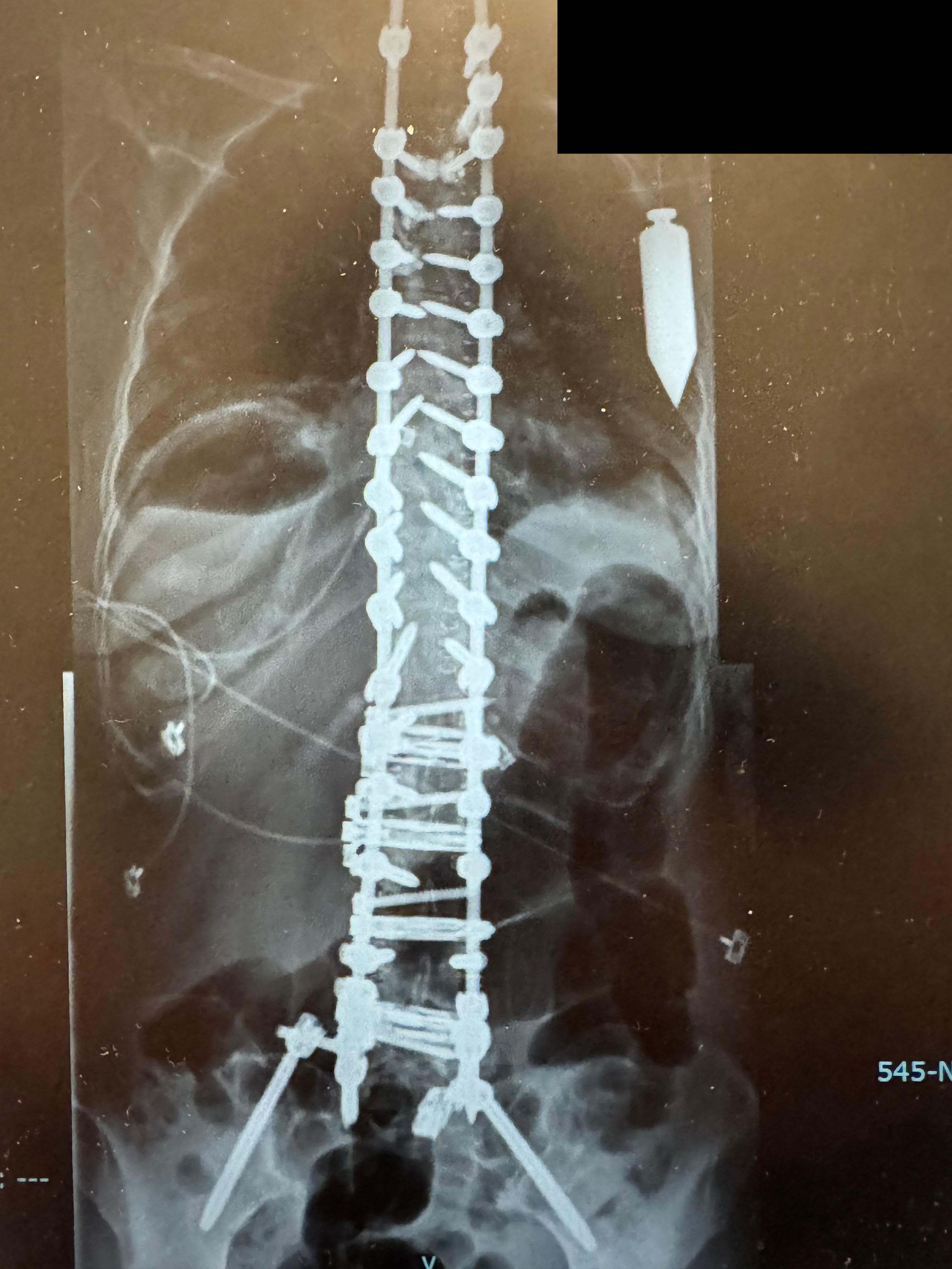
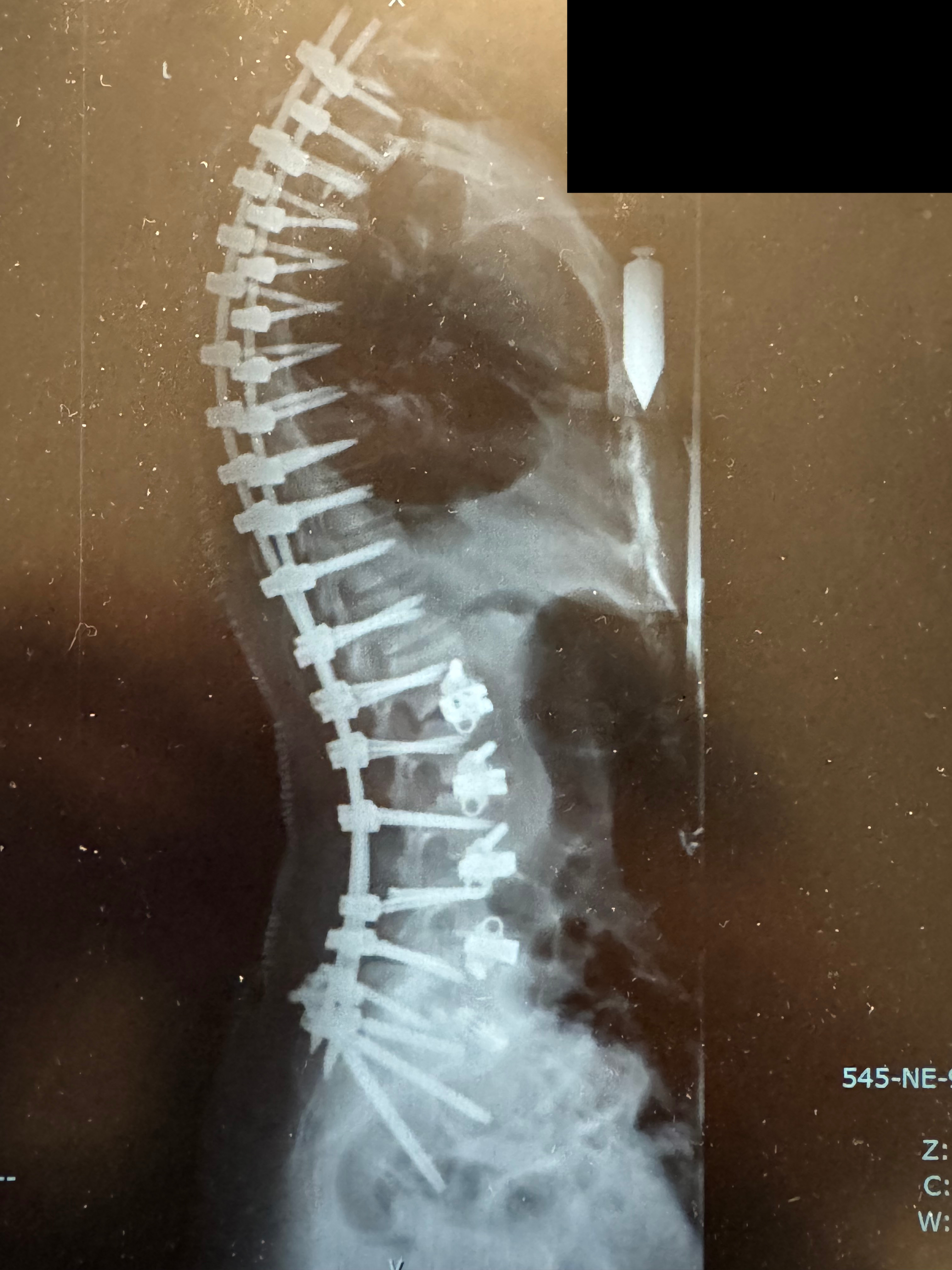
Post-Operative Imaging

Post-operative AP X-ray showing corrected alignment with instrumentation
AP View: Lateral curve corrected with bilateral screws and rods providing stability
Post-operative lateral X-ray showing restored sagittal balance
Lateral View: Kyphosis corrected, sagittal balance restored, upright posture achieved

Surgical Approach

Procedure: Multi-level posterior spinal fusion with pedicle screw instrumentation from T12 through L5

Technique: Open posterior approach with decompression, osteotomy for kyphosis correction, and fusion with bone graft

Outcome: Complete correction of both scoliotic curve and kyphotic deformity with restored sagittal balance and upright posture. Patient reports excellent pain relief and restoration of walking tolerance.

What to Expect After Surgery

Recovery Timeline

Recovery from adult scoliosis surgery follows a predictable pattern. Most patients experience significant pain relief and improved function within the first 3-6 months.

Day 1–3
Hospital Stay
Most patients stay 2-3 days. Pain is well-controlled with medication. Physical therapy begins immediately with walking and basic movements.
Week 1–6
Early Recovery at Home
Rest with gradual increase in activity. Avoid heavy lifting and twisting. Most patients notice improved posture within weeks. Pain medication typically reduced significantly.
Week 6–12
Active Rehabilitation
Formal physiotherapy. Core strengthening and balance training. Most patients return to office work and light activities. Walking distance increases progressively.
Month 3–6
Functional Improvement
Most patients report significant improvement in pain, walking and daily function. Return to moderate activities like golf, gardening and gym work. Neurological symptoms often resolve.
Month 6–12
Bone Fusion and Stability
Fusion solidifies. Most restrictions lifted. Return to all normal activities. Continued improvement in posture, balance and leg symptoms. Maximum functional recovery achieved.

Recovery after scoliosis fusion is gratifying. Most patients experience dramatic relief from pain and restoration of the ability to stand upright and walk normally. The physical transformation — straightening of the spine and restoration of balance — is often visible within weeks.

The most important predictor of outcome is choosing the right surgical approach for your specific deformity. Multi-level fusion is a major procedure, but modern techniques and minimally invasive options have made it safer and recovery faster than ever.

Fusion is permanent. The fused levels no longer move, but the remaining spine continues to function normally. Regular activity and core strengthening help maintain long-term results.

Ready to Get Better?

Talk to Dr. Ganapathy About Your Scoliosis

If you have been told you have scoliosis or are experiencing progressive back pain and imbalance, a comprehensive evaluation can determine the best path forward.

Book a Consultation
Arizona Locations Coming Soon

How we think through this condition

Every spine decision should answer the same practical questions before surgery is considered.

What is causing the symptoms?We connect your pain, weakness, numbness or walking limits to imaging and exam findings.
What has already been tried?Physical therapy, medications, injections and time matter when they fit the diagnosis.
What are the surgical goals?The goal may be nerve relief, stability, deformity correction or preserving function.
What is the recovery plan?You should know the expected timeline, restrictions and support needs before making a decision.