Advanced surgical techniques for excessive thoracic spine curvature. Restore your posture, mobility, and quality of life with expert spine surgery.
Kyphosis is an excessive forward curvature of the thoracic spine (mid-back), creating what's commonly called a "dowager's hump" or stooped posture. While a normal amount of thoracic kyphosis is healthy, excessive curvature can significantly impact your mobility, breathing, and quality of life.
Kyphosis develops from multiple causes including osteoporosis, degenerative disc disease, spinal fractures, poor posture over decades, or structural abnormalities. Age-related changes are common, but younger patients can develop kyphosis from trauma or underlying conditions.
Beyond cosmetic concerns, untreated kyphosis can lead to chronic pain, breathing difficulties, balance problems, and reduced ability to look upward. Early diagnosis and treatment options range from physical therapy to advanced surgical correction depending on severity.
Unable to maintain upright posture; chin droops toward chest making eye contact difficult.
Visible rounded hump at the base of neck; significant forward curvature of upper back.
Persistent pain in mid-back and neck from muscle strain and vertebral stress.
Shortness of breath during normal activities due to compressed chest cavity.
Difficulty maintaining balance; increased risk of falls and related injuries.
Cannot comfortably look upward or at ceiling; limited neck extension.
Assessment of posture, spinal curve, and mobility. Neurological testing to check for nerve involvement and assess pain distribution.
X-rays to measure Cobb angle (degree of curvature). MRI or CT scans to evaluate disc health, bone density, and nerve compression.
Standard measurement that determines kyphosis severity and treatment recommendations. Essential for surgical planning and outcome assessment.
Testing breathing capacity, balance, and daily activity limitations. Helps determine surgical urgency and expected improvement.
Multiple vertebrae are fused together using rods, screws, and bone grafts to stabilize the spine and correct excessive curvature. Provides long-term correction and prevents progression.
For kyphosis caused by osteoporotic fractures, specialized techniques restore vertebral height and alignment, reducing pain and preventing further collapse.
Minimally invasive procedure that uses a balloon to restore vertebral body height in compression fractures, followed by cement injection for stabilization.
For severe fixed deformities, controlled bone cuts and realignment restore normal posture. Advanced technique requiring specialized expertise.
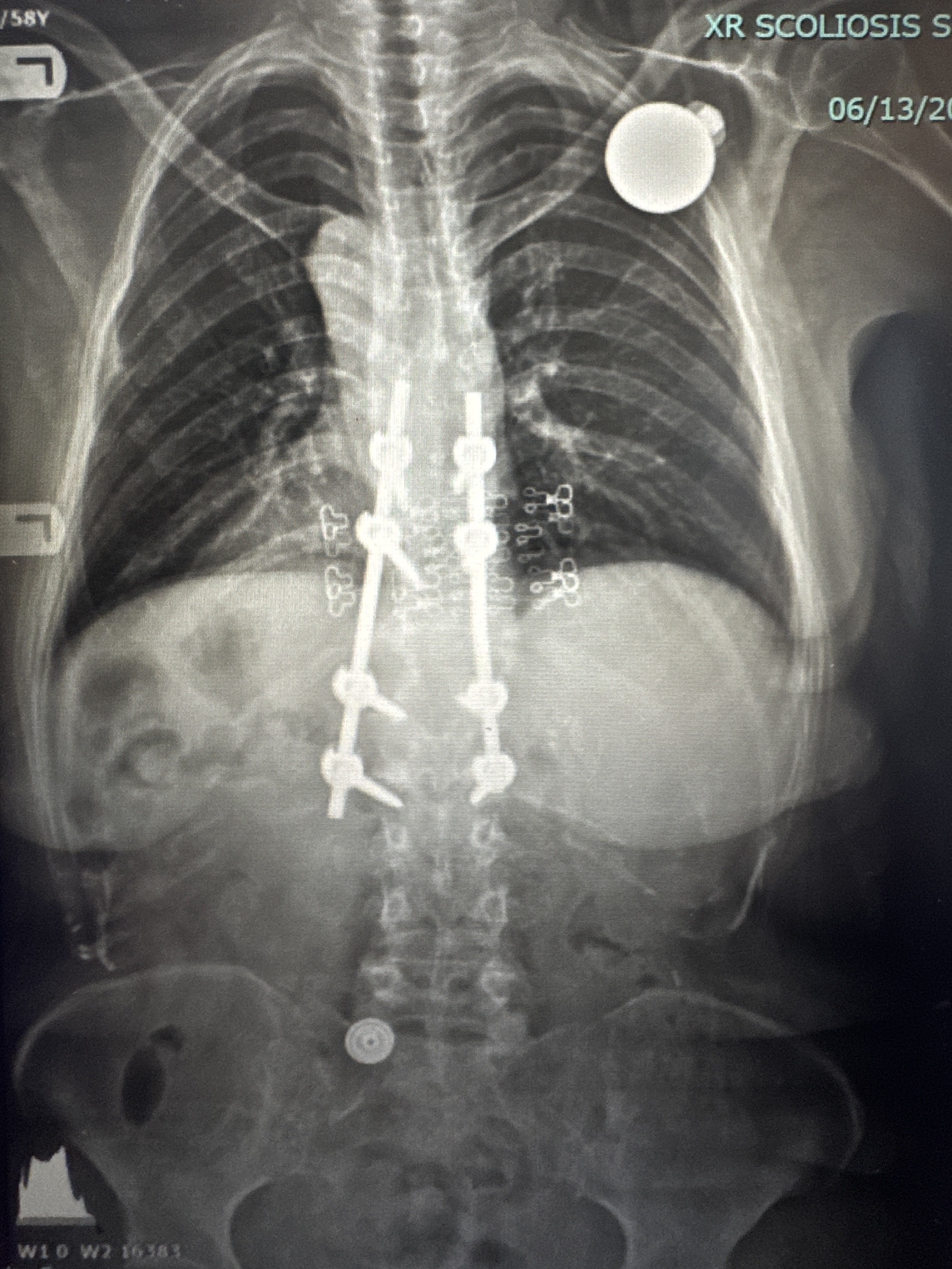
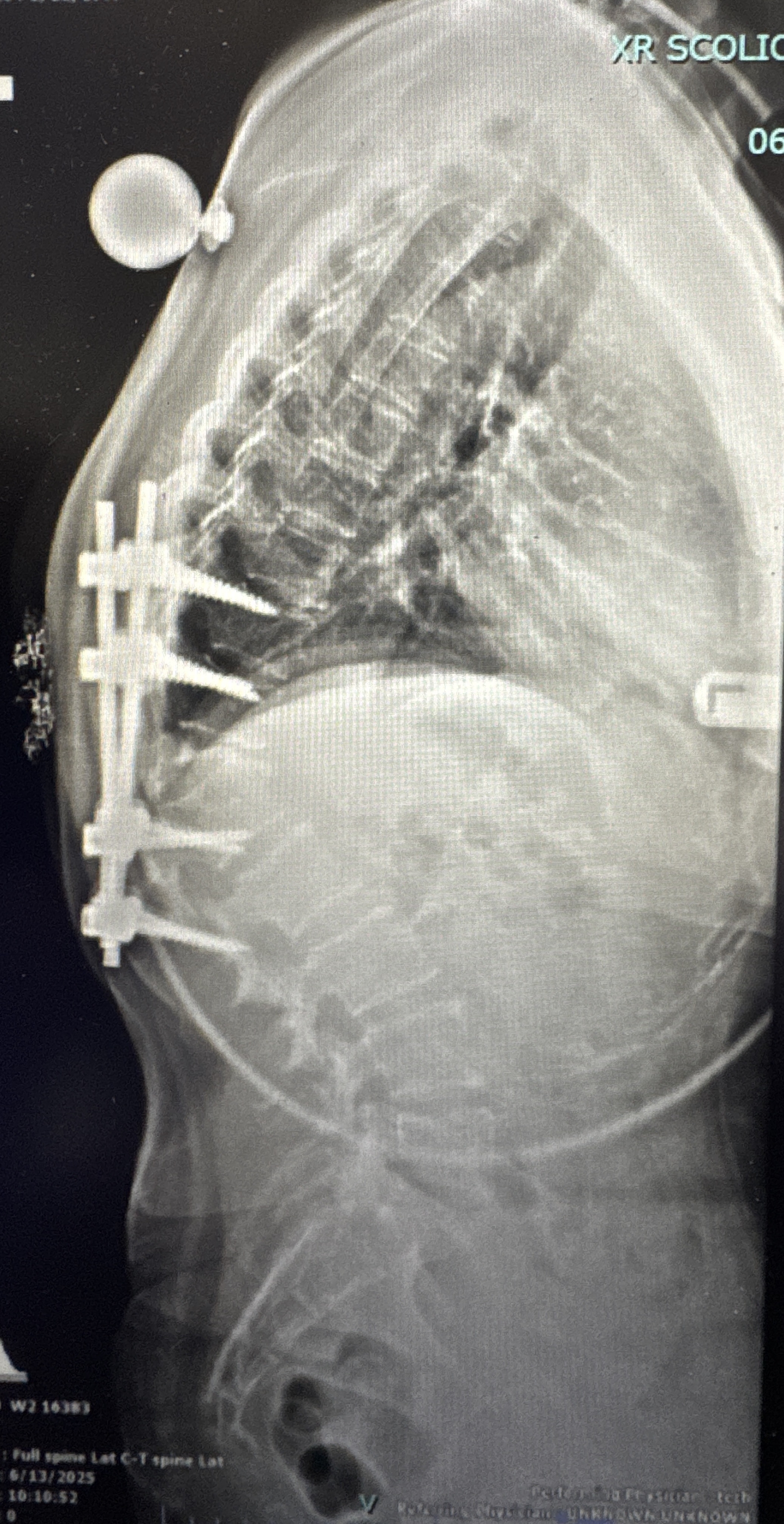
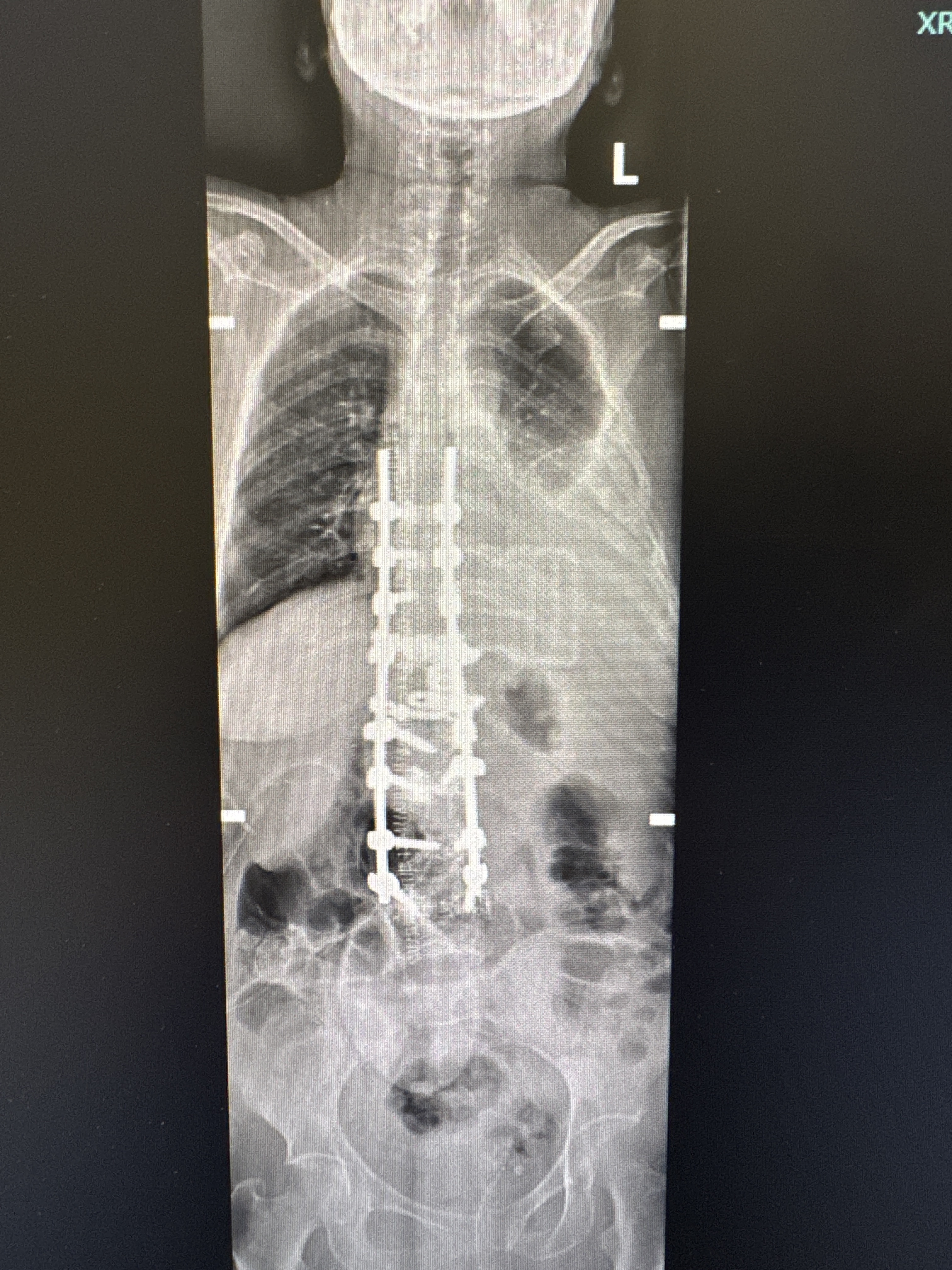
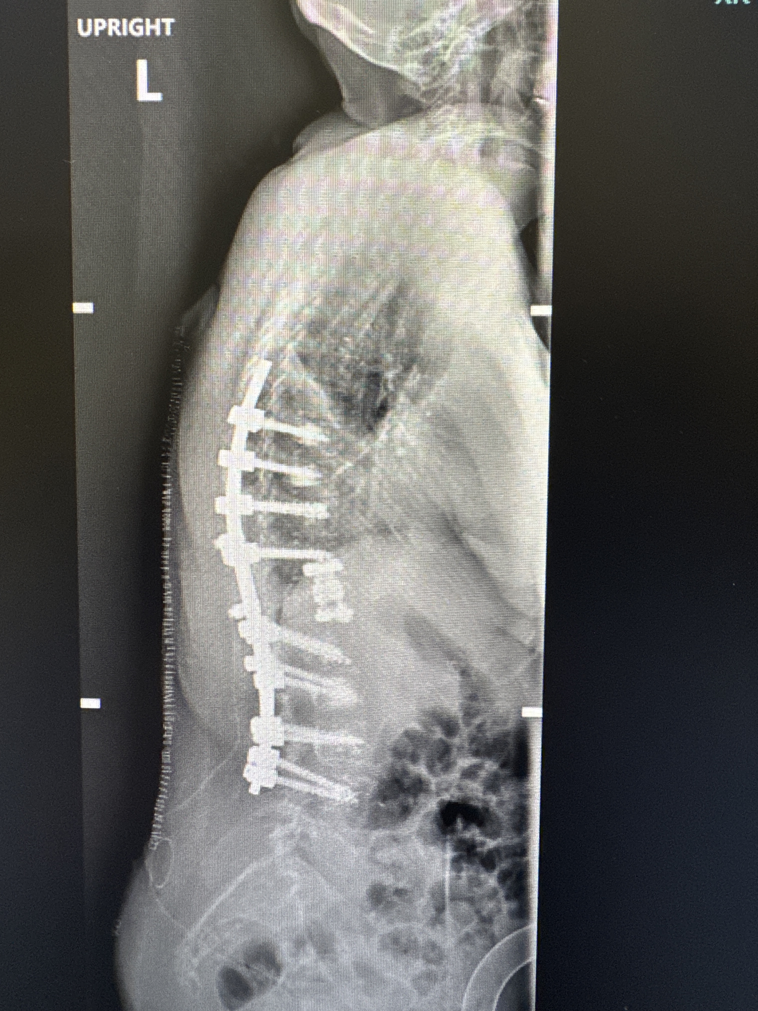
Real patient case showing successful correction of combined thoracolumbar scoliosis and kyphotic deformity using multi-level posterior fusion with instrumentation.
Procedure: Multi-level posterior spinal fusion with pedicle screw instrumentation from T12 through L5
Technique: Open posterior approach with decompression, osteotomy for kyphosis correction, and fusion with bone graft
Outcome: Complete correction of both scoliotic curve and kyphotic deformity with restored sagittal balance and upright posture. Patient reports excellent pain relief and restoration of walking tolerance.
Schedule a consultation with Dr. Venkat Ganapathy to discuss your treatment options and create a personalized care plan.
Schedule ConsultationEvery spine decision should answer the same practical questions before surgery is considered.